Radiology
Radiology - ACL Post Surgical Complications

Sport and Exercise Physician | Managing Director
Published
March 2, 2026
Sport and Exercise Physician | Managing Director
Published
March 2, 2026
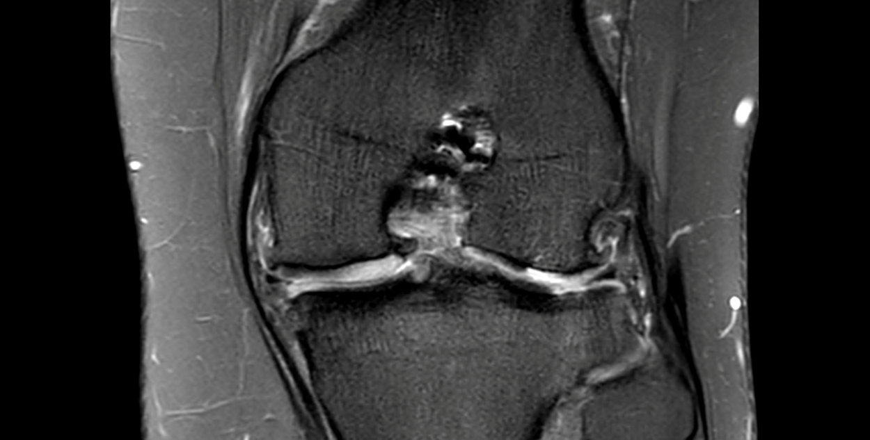
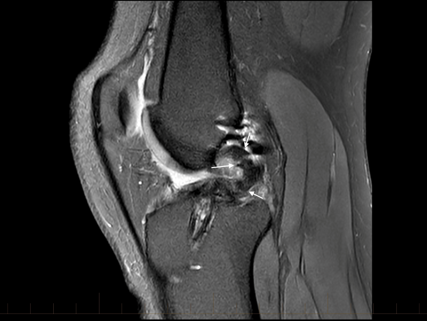
What post-surgical complications, following an ACL reconstruction, are shown on this coronal MRI sequence?
A 21-year-old female player presented after a twiring injury to her left knee. She describes a feeling of instability in her knee, rapid swelling and an inability to continue playing. On examination she had gross laxity with Lachman Test, an effusion and a restriction ROM.
The player had a history of a previous ACL reconstruction (ACLR). This was done using hamstring autograft two years previously. At the time of this surgery (done on another country) her operation note reports that she had both medial and lateral meniscal repairs. She recovered well from this surgery and had been playing elite football for more than one year.
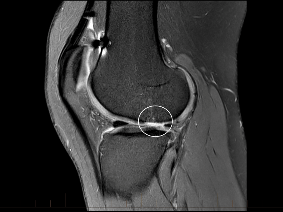
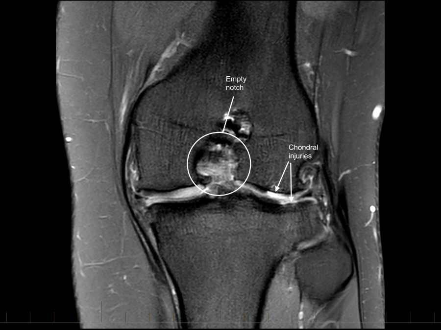
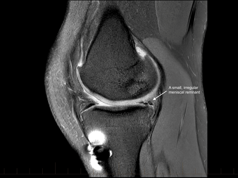
The ACL graft is ruptured with a loss of the normal ACL contour and signal. There is a focal are of full-thickness cartilage loss on the posterior, weight-bearing portion of the lateral femoral condyle. This measure approximately 15 x 10mm. There is also thinning and irregularity of the articular cartilage involving the posterior aspect of the lateral tibial plateau with a small area of full thickness cartilage loss and subchondral reactive change. These chondral changes were not present at the time of her first surgery. There appears to have been trimming of the mid and posterior thirds of both menisci. These meniscal remnants are small and irregular.
This player opted to have a revision ACLR. This was done using bone-patella-bone autograft and augmented with an ALL tenodesis. She also had a chondroplasty and further partial medial meniscectomy. She is currently three months post-surgery and is progressing well with her rehabilitation.
This case highlights several important points related to ACL injuries.
Firstly, there is a high rate of re-injury. Data from various registries and cohort studies suggests that the risk of sustaining a further ACL injury (either to the injured or contralateral knees) is approximately 30% for young, active patients who return to change of direction sports like football.
Secondly, there is a high incidence of further chondral and meniscal injuries as well as post-traumatic osteoarthritis. Despite the initial surgeon's attempts to repair the meniscal injuries after this player's first ACL injury, she has gone on to develop significant chondral pathology. This has occurred over a relatively short period.
Finally, this case re-enforces the need to advocate ACL prevention programmes. Despite this player's high risk for re-injury, she was never told about prevention programmes, like the 11+. When done regularly these programmes have been shown to reduce the risk of ACL injury by approximately 50%. Given that these programmes are likely more effective in higher risk populations, the value to this particular player may have been even greater.