


Radiology
Radiology Review - More Than an ACJ Sprain.

Sport and Exercise Physician | Managing Director
Published
October 1, 2026
Sport and Exercise Physician | Managing Director
Published
October 1, 2026
.png)
Persistent or recurring ACJ pain after a shoulder injury is not always another sprain. In this case, a 23-year-old rugby league player presented with pain in his dominant right shoulder. The pain was well localised to the acromioclavicular (ACJ) region, with focal tenderness on examination. He had sustained an ACJ sprain approximately one year earlier. His symptoms initially settled and he returned to play. However, the pain subseqently recurred, coinciding with an increase in upper-body strength training with his team. He had been diagnosed with a new 'sprain'.
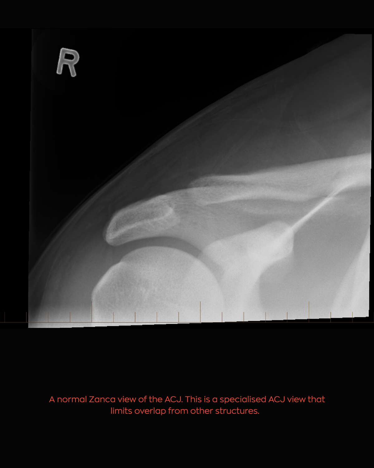
X-rays taken at the time of his original injury were normal. With his recurrent symptoms, repeat X-rays were obtained.
The original 'Zanca' view of the right shoulder was normal.
Repeat X-rays taken approximately one year later demonstrated a clear interval change. There is irregularity and bone resorption involving the distal end of the right clavicle. The adjacent acromion was also mildly irregular, and the acromioclavicular joint is widened. The coracoclavicular interval remained normal. These appearances are consistent with distal clavicle osteolysis.
Distal clavicle osteolysis is an important differential diagnosis in an athlete with persistent or recurrent pain following an ACJ injury. It may occur following trauma or in association with repetitive microtrauma. In athletes, heavy upper body resistance training, particularly repetitive pressing exercises such as bench press, military press and push-ups, can place considerable load across the ACJ.
In this case, the initial images were normal. The development of clear radiographic abnormalities over the following year highlights an important clinical point, consider distal clavicle osteolysis when symptoms following an ACJ sprain do not settle as expected, or when ACJ pain recurs after an initial period of improvement. X-rays can be very helpful in establishing the diagnosis and may be the only imaging required. Typical findings include osteopenia and resorption involving the distal clavicle, often with loss of irregularity of the articular cortical margin. If further investigation is required, MRI is generally the imaging modality of choice. Increased T2 signal around the distal clavicle and ACJ is a common finding. A diagnostic local anaesthetic injection into the ACJ can also be useful when the source of symptoms remains uncertain.
This player was managed with education, a short course of oral NSAIDs, physiotherapy and relative rest.
He was able to continue rugby league training and playing while modifying his upper body strength programme to avoid provocative pressing exercises. His symptoms gradually settled over approximately three months. Treatment of distal clavicle osteolysis is generally conservative initially, with activity modification and time. Other options include corticosteroid injection and, in persistent cases, surgical excision of the distal calvicle.
When ACJ pain fails to settle, or settles and then returns, consider distal clavicle osteolysis, particularly in athletes undertaking significant upper body resistance training.
.png)


