Radiology
Radiology Review - Brachial Neuritis Suprascapular Nerve Spine

Sport and Exercise Physician | Managing Director
Published
August 4, 2026
Sport and Exercise Physician | Managing Director
Published
August 4, 2026

Shoulder weakness is often linked to rotator cuff injury or structured pathology, but nerve-related causes should also be considered.
This case highlights suprascapular neuritis presenting with marked weakness and muscle wasting following a brief episode of severe shoulder pain, with MRI playing an important role in identifying muscle denervation and excluding mechanical compression.
Severe pain followed by persistent weakness
A 45-year old male patient presented with a six-month history of left shoulder weakness. At the time of his visit, he did not have any significant shoulder pain (or other symptoms).
Talking with him however he did report experiencing a ten-day period, immediately prior to developing his current symptoms, where he had experienced severe pain in his shoulder.
On examination there was visible wasting within the infraspinous fossa and significant weakness of external rotation.
There was mild weakness of resisted supraspinatus testing (empty can) but no clear wasting. His shoulder examination was otherwise normal.
There was a normal spin examination and no other focal neurological signs.
MRI evidence of muscle denervation
An X-ray of the shoulder was normal.
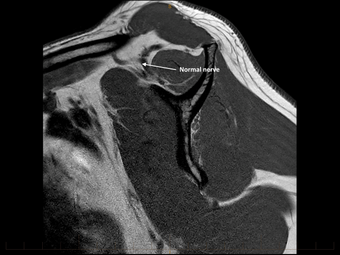
MR images demonstrate atrophy and marked oedema within the supraspinatus and infraspinatus muscle bellies. There is no fatty replacement.
The remainder of the rotator cuff musculature, deltoid and the partially visualised trapezius are normal in signal.
Perineural fat planes are maintained around the suprascapular nerve within the infraspinatus fossa, spinoglenoid notch and suprascapular notch. The nerve appears anatomically normal.
There is no evidence of mechanical compression.
A characteristic presentation of brachial neuritis
This patient was diagnosed with brachial neuritis (Parsonage-Turner syndrome)
The sudden onset of short-lived, sever shoulder pain followed by weakness is a characteristic history for this problem.
The absence of clear trauma, history of repeated overhead activity or a structural lesion on MR imaging also make this diagnosis more likely.
He has been referred for nerve conduction studies / EMG to help confirm the distribution of nerves affected, and to provide an indication about nerve reinnervation and prognosis.
He has been advised that spontaneous recovery is the most likely outcome however this may take up to 36 months.
The role of MRI and nerve conduction studies
Muscle denervation has multiple possible causes, including trauma, neuropathies, infection and neoplasia.
MRI is a very useful study as it can both diagnose muscle denervation and help determine why this has occurred (for example by illustrating a structural lesion).
MRI demonstrates characteristic signal intensity patterns depending on the chronicity of muscle denervation.
In the acute or sub-acute phases, as illustrated by this case, denervation muscle shows a high signal intensity pattern on fluid sensitive sequences and normal signal intensity on T1-weighted MRI images.
In chronic denervation, muscle atrophy and fatty infiltration can be seen. This is generally best seen on T1-weighted sequences with high signal and volume loss.
Nerve conductions studies and EMG are often done alongside MR imaging.
These tests can help localise areas of compression or neuropathy and can provide information and functional recovery.