Radiology
Radiology Review - Hamstring Muscle Injury

Sport and Exercise Physician | Managing Director
Published
September 1, 2026
Sport and Exercise Physician | Managing Director
Published
September 1, 2026
Hamstring injuries are common in running and field-based sports, particularly during high-speed sprinting. While clinical assessment remains central to diagnosis and rehabilitation planning, MRI can help define the location and extent of injury and provide a more detailed atomical classification.
This case highlights how imaging findings can appear extensive despite relatively modest clinical symptoms, and why MRI classification should be interpreted alongside the athlete's clinical presentation and functional progress.
A 25-year-old male professional footballer developed sudden pain and tightness in his left posterior thigh while accelerating into a maximal sprint. He stopped training immediately and was assessed the following day.
He walked normally but reported discomfort with a longer stride and faster walking. Examination demonstrated localised tenderness over the lateral hamstring, awareness with straight-leg raises and mild pain with MHFKE testing. There was no major loss of power, bruising or a palpable defect.
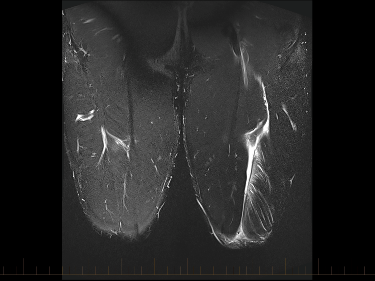
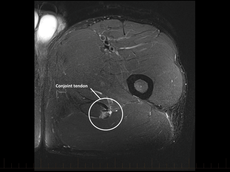
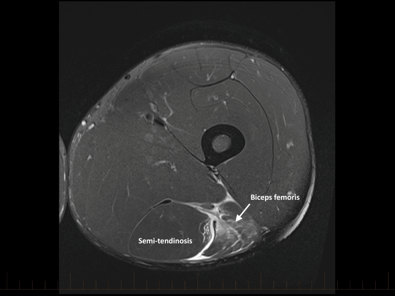
MRI demonstrates extensive oedema throughout the long head of biceps femoris, extending from the proximal region to approximately 10cm above the knee. The signal abnormality is centred on the main musculotendinous junction and radiates through the muscle belly.
There is a mild distortion of the myotendinous fibres but no significant fibre gap, retraction or loss of tension. Haemorrhage extends along the intermuscular septum, with minor involvement of the lateral semitendinosus disruption.
Because the oedema extends for more than 15cm and is centred on the musculotendinous junction, the principal injury is classified as BAMIC 3b. The suffix 'b' denotes a musculotendinous injury; the grade is driven by the extensive craniocaudal length of signal abnormality rather than by a large fibre defect.
The British Athletics Muscle Injury Classification (BAMIC) grades muscle injuries from 0 to 4 according to their MRI extent and adds a suffix describing the principal anatomical site: 'a' for myofascial. 'b' for musculotendinous and 'c' for intratendinous injury.
This provides a more precise description than the traditional grade 1-3 terminology and draws attention to intratendinous injuries, which may require a more cautious rehabilitation. In this case, a long area of oedema produces a BAMIC 3b classification despite limited fibre disruption and relatively modest clinical findings.
BAMIC is useful for describing where an injury is located and how extensive it appears, but it should not be treated as a return-to-play calculator. Some cohort studies have found associations between higher BAMIC grades, intratendinous involvement and longer time to return.
However, findings vary between sports and studies, and there is substantial overlap in recovery time between grades. Systematic reviews have found no strong evidence that an individual MRI feature reliably predicts return-to-play time or reinjury. MRI also adds little prognostic information beyond a careful history and clinical examination in many acute hamstring injuries.
Abnormal signal commonly persists after clinical recovery. Return-to-play decisions should therefore be based on serial clinical assessment and successful progression through strength, running, sprinting and football-specific rehabilitation, rather than repeat imaging or the BAMIC grade alone.
This player completed a criteria-based rehabilitation programme and returned to competitive football after 19 days without recurrence. His progression was determined clinically rather than by the extensive MRI signal abnormality.